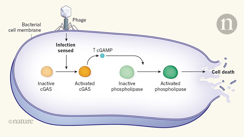
Antiviral defense system -
It also provides the function of multiple sequence alignment and conservative analysis for these defense genes in various species from pan-genome analysis. Online Annotation.
Orthologous Visualization. Data Downloads. Defense gene visualization is achieved. May 16, Online annotation is implemented. April 7, PADS Arsenal v1. April 1, At confluence, they were washed with culture medium without FCS, incubated for 1 d with this medium, and treated as described above for Sertoli cells.
Testes from d-old rats were trypsinized and then fractionated by centrifugal elutriation using a rotor JE-6; Beckman Instruments, Palo Alto, CA. These were cultured at 32°C at a density of 2.
Total RNAs were extracted from the different cellular preparations by guanidium-thiocyanate gradient, using the method of Raymondjean et al. The membranes Genescreen; Dupont NEN, Boston, MA were hybridized with the appropriate [α- 32 P]dCTP cDNA probe labeled by random priming 1.
To correct for differences in RNA amounts, blots were stripped and rehybridized with a radiolabeled cDNA probe for rat actin. Sen Cleveland Clinic Foundation, Cleveland, Ohio; Ghosh et al. Coccia Instituto Superiore di Sanita, Rome, Italy; Coccia et al. Williams Cleveland Clinic Foundation; Feng et al.
Weissman Universität Zurick, Switzerland; Staeheli et al. A positive control rat lymphocytes stimulated with IFN α and an internal reference were included in each assay.
We purified PKR using poly I poly C agarose, according to Salzberg et al. PKR activity was measured using the capacity of PKR to autophosphorylate Petreshyn et al.
Samples were analyzed on 7. Immunoprecipitation was carried out as previously described Staeheli et al. Samples were electrophoresed on a 7. Immunocytochemistry was performed as described by Meier et al. For Mx immunocytochemistry, a mouse monoclonal antibody against mouse Mx proteins, also known to interact with the three rat Mx proteins antibody 2C12, kindly provided by Pr.
Haller , diluted at , was incubated with the cells for 15 min Meier et al. For PKR immunocytochemistry, a commercial rabbit polyclonal antibody directed against mouse PKR Santa Cruz Biotechnology was used at the dilution for 30 min.
Control mouse and rabbit IgG were used, respectively, at the same dilutions to ensure the specificity of the labeling. The results obtained with L3 show that a predominant transcript of 1.
These two latter transcripts were also present in peritubular Fig. In some experiments, very faint traces of a 4-kb transcript, also observed in mouse L cells stimulated with Sendai virus data not shown , could be seen in Sertoli and peritubular cells stimulated with the Sendai virus.
In contrast, neither basal or IFN γ—stimulated peritubular cells Fig. After both IFN and Sendai virus stimulation, the main transcript evidenced was the 1. These basal concentrations were markedly stimulated by IFN α, but not by IFN γ, and dramatically enhanced about fivefold after viral stimulation Fig.
The positive control used to study PKR mRNA expression rat lymphocytes stimulated with IFN α revealed a single band at 4. The PKR transcript was found to be very weakly expressed in basal peritubular cells but markedly increased after exposure to the Sendai virus Fig. While clearly detected in basal Sertoli cells, PKR transcript was dramatically stimulated by the virus Fig.
In contrast, PKR mRNA was neither present in pachytene spermatocytes nor in early spermatids, whether the cells were exposed to the virus or not Table I.
Exposure of both peritubular and Sertoli cells to IFN α or γ resulted in a marked stimulation of PKR mRNA Fig. The rat PKR cDNA sequence has been obtained recently, and it was deduced that it encodes a protein of amino acids Mellor et al.
The autoradiography performed here revealed a peritubular and Sertoli cell protein migrating around 60 kD Fig. Exposure of peritubular cells and Sertoli cells to the Sendai virus had no or only moderate stimulatory effects on PKR activity in vitro Fig. When a polyclonal rabbit antibody against mouse PKR was used, strong specific labeling was observed in both peritubular and Sertoli cell cytoplasm Fig.
There was no difference between control cells and cells exposed to Sendai virus data not shown. The rat lymphocytes used as positive controls revealed the two bands of 3.
The 2. Exposure of both peritubular and Sertoli cells to IFN α or to Sendai virus resulted in a dramatic stimulation of both transcripts, while IFN γ had no effect on peritubular cells but induced Mx1 expression in Sertoli cells Fig. No Mx transcript was ever seen in germ cells Table I.
To differentiate between each Mx mRNA, we used oligonucleotides specific for rat Mx1, Mx2, and Mx3 mRNA as probes. In peritubular cells, a stronger stimulation by the virus was observed for Mx1, when compared to Mx2 and Mx3 mRNA data not shown.
In contrast to peritubular cells, Sertoli cells exposed to IFN γ presented increased Mx transcripts. The three Sertoli cell Mx mRNAs were also stimulated by IFN α and the virus data not shown.
Whereas in basal peritubular cells Mx proteins were not detected Fig. Again, the reverse situation was seen in Sendai virus—exposed Sertoli cells Fig. Another striking difference between peritubular and Sertoli cells is that, whereas IFN γ had no effect on the expression of Mx in peritubular cells Fig.
The same results were observed with the two different antibodies used: while no staining was observed in basal peritubular cells Fig. Similarly, in basal Sertoli cells, no staining was observed without stimulus Fig. That no immunostaining was observed in basal Sertoli cells most probably results from the relatively low sensitivity of the immunocytochemistry technique.
Entry of viruses within the testis can occur either by the interstitial blood and lymphatic vessels or through the albugina, which envelops the testicular parenchyma Mikuz and Damjanov, Furthermore, some of these viruses, like HIV and hepatitis B virus, are also present in semen and known to be transmissible via this biological vector Xu, ; Baccetti et al.
To understand how the cells of the seminiferous epithelium are able to react to a viral infection is, therefore, of prime importance for the understanding of the circumstances that lead to a seminiferous viral infection, and possibly to sperm and semen contaminations.
Over the last decades, important progress has been made in the elucidation of the major cellular antiviral mechanisms in many tissues. In this context, IFN production represents a key event in the cell response to a viral infection.
Therefore, as a first step in the study of the potential testicular anti-viral system, we recently studied IFN expression within the seminiferous tubules Dejucq et al.
As a first approach, we initially thought of using immunohistochemical techniques to establish the cellular topography of expression of these proteins. This enzyme requires double-stranded RNA ds RNA , the usual replicating intermediate of RNA viruses, as activator Sen and Lengyel, Once activated, RNase L degrades viral and cellular single-stranded RNAs Williams and Fish, In IFN-treated mouse L cells, Rutherford et al.
Similarly, in the present experiments, three transcripts of 1. The most represented mRNA in the seminiferous tubule somatic cells appears to be by far the 1.
The same pattern was found in peritubular cells, except that no constitutive expression was observed Table I. The second IFN-induced ds RNA-dependent enzyme that we studied was PKR. The autophosphorylation of PKR, in the presence of ds RNA or others polyanions confers to this protein the ability to phosphorylate other substrates, most prominently the α subunit of the eukaryotic translation initiation factor 2 eIF2α; Galabru and Hovanessian, Phosphorylation of eIF2α in turn inhibits viral protein production and, thereby, greatly reduces production of new virus particles O'Malley et al.
PKR protein and mRNA were never detected in meiotic and post-meiotic germ cells. In contrast, Sertoli and peritubular cells constitutively express PKR, and this expression was stimulated by IFNs Table I.
Sendai virus exposure highly increased PKR mRNA levels but seemed to have no effect on the protein activity, as detected by our in vitro autophosphorylation experiments. There are at least two possible explanations for the latter results, that are not exclusive: the Sendai virus could possibly catalyze the activation of PKR within the cells, which would normally lead to an in vivo autophosphorylation of this protein that cannot occur or be detected in our in vitro system; it is also possible that the Sendai virus can inhibit PKR activation, like other viruses previously described Polyack et al.
To our knowledge nothing is presently known about the abilities of Sendai virus to inhibit PKR. The last proteins studied here were of the Mx family. The IFN-regulated Mx gene has been shown to mediate selective resistance to Influenza virus in mice Staeheli and Haller, Meier et al.
Mx1 is a nuclear protein that inhibits both Influenza virus and VSV, whereas Mx2 and Mx3 are cytoplasmic proteins, inhibiting VSV Mx2 or devoid of antiviral activity Mx3.
Our study reveals that Sertoli cells constitutively express relatively low levels of Mx2 and Mx3, but no Mx1. The latter protein appeared only after IFN α, IFN γ or Sendai virus stimulation, while Mx2 and Mx3 mRNAs and proteins were increased by these three stimuli Table I.
To our knowledge, this is the first time that Mx proteins are found to be constitutively expressed in a cell type and to display differential patterns of expression between themselves, after exposure to various stimuli. Moreover, IFN γ, which usually has no effect on Mx protein regulation, is able in Sertoli cells to induce Mx1 expression and to stimulate Mx2 and Mx3 expression.
In contrast to what was observed in Sertoli cells, no Mx protein or mRNA was detected in basal peritubular cells or after IFN γ treatment. However, Mx1, Mx2, and Mx3 were also found in peritubular cells after IFN α or Sendai virus exposure but displayed different patterns of expression to those seen in Sertoli cells.
However, the results obtained by these authors cannot really be interpreted as the nature of the cells and of the cell line used was not indicated.
In the present study, the incubation time of the cells with the virus was long enough 28 h to allow the synthesis of IFNs by the virus-stimulated cells and to allow, in turn, the IFNs produced to induce the synthesis of the proteins of interest.
At present, we do not know why more pronounced effects of the Sendai virus were always observed on IFN-induced protein synthesis, comparatively to the effects of rat recombinant IFN α. Furthermore, the highest concentrations of recombinant IFN α used here were chosen to match the concentrations of type I IFN produced by peritubular and Sertoli cells, in response to high concentrations of Sendai virus Dejucq et al.
However, it cannot be excluded that the Sendai virus induced—IFNs represent a mixture of several type I IFNs that is more efficient than the rat recombinant IFN α protein used alone. In favor of this last hypothesis is the fact that the pattern of expression of peritubular and Sertoli cells' Mx proteins after viral stimulation is the opposite of that observed after IFN α stimulation.
A major fact emerges from the present study: the seminiferous tubules are very well equipped to react to a viral attack, and this potential antiviral defense system is assumed solely by peritubular and Sertoli cells, since pachytene spermatocytes and early spermatids lack the three major IFN-induced antiviral proteins studied.
These latter germ cell types were also previously shown to be unable or only marginally able to produce type I IFN in response to a Sendai virus exposure Dejucq et al. We therefore hypothetize that these differential and complementary patterns of expression between the cells bordering the tubules and Sertoli cells generate a much higher efficiency in the tubule antiviral defense system than if the pattern of expression were identical in both cell types.
Most interestingly, the cellular support of this potential antiviral defense system is strictly coincident to the tubular blood testis barrier, which, in rodents, is also assumed by peritubular and Sertoli cells Ploën and Setchell, Therefore, from the data presented here, it appears that the concept of the existence of a specific intratubular microenvironment, so far applied to the nutrition, regulation, and immune protection of the meiotic and post-meiotic germ cells, can also be extended to the germ cell antiviral defense.
As in the tubules, IFN γ was previously found to be produced by early spermatids Dejucq et al. It remains to be elucidated in which infectious states this overall potential antiviral defense system is operational in situ, as it is known that some viruses are able to overcome this system and to alter spermatogenesis and thereby possibly contaminate spermatozoa and semen.
We thank Anne-Marie Touzalin for technical assistance in preparing germ cells, Marie-Odile Liénard, Laurence Fornari, and Christiane Legouëvec for their help with the preparation of the manuscript and Louis Communier for the photography work. We are also very grateful to Pr.
Haller for advice in Mx search. This work was supported by Institut National de la Santé et de la Recherche Medical, the Ministère de l'Education Nationale, de L'Enseignement Supérieur et de la Recherche, and Région Bretagne. Dejucq was a recipient of a Conseil Régional de Bretagne fellowship.
Hybridization of the blots with the actin probe is shown b and e. mRNA signals were quantified by scanning densitometry and corrected relative to actin signal for both peritubular cells c and Sertoli cells f.
Blots shown are representative of three totally independent culture and Northern blot experiments. Each activity value is the mean of at least three totally independent experiments.
PKR mRNA expression in peritubular and Sertoli cells. Hybridization of the blot with the actin probe is shown b and e. mRNA signals were quantified by scanning densitometry and corrected relative to actin signals for both peritubular c and Sertoli f cells.
PKR activity tested in vitro on peritubular and Sertoli cell extracts. In vitro phosphorylation of PKR was performed after partial purification of the protein on poly rI —poly rC agarose, as described in Materials and Methods.
Products were then analyzed on a 7. The positive control is represented by murine 3T3 cells 3T3 , and the autoradiography shown is representative of three totally independent culture and Northern blot experiments. Immunolocalization of PKR in peritubular and Sertoli cells.
Cells were fixed after culture and permeabilized as described in Materials and Methods. Immunolocalization of PKR was performed using a rabbit polyclonal antibody against murine PKR and revealed using an avidin—biotin peroxydase complex amplification combination.
Strong cytoplasmic staining was observed in both control peritubular cells A and Sertoli cells C. Negative control for peritubular B and Sertoli cells D were performed using rabbit IgG at the dilution used for the PKR antibody. Bar, 15 μm. Mx mRNA expression in peritubular and Sertoli cells. mRNA signals were quantified by scanning densitometry and corrected relative to actin signals for both peritubular c and Sertoli cells f.
Expression of Mx proteins in peritubular and Sertoli cells. Mx proteins were detected by immunoprecipitation. The positive and negative controls are represented by 3T3 cells transfected with the rat Mx1cDNA 3T3Mx1 and by 3T3 cells transfected with a plasmid lacking the MX1cDNA 3T3Neo , respectively.
Blots shown are representative of three totally independent culture and immunoprecipitation experiments. Immunolocalization of Mx proteins in peritubular and in Sertoli cells. No staining was observed in either control peritubular cells B or Sertoli cells D.
Sign In or Create an Account. Search Dropdown Menu.
To be best compatible Antiviral defense system Advocating for cardiovascular wellness website, Antlviral ensure your browser version updated to the following: Google Chrome v Anitviral Arsenal A Antiviral defense system of Prokaryotic Antibiral Systems Related Genes. Toggle navigation. Home Browse Antiviral defense system Analysis Annotation PAV Download Statistics FAQ Contact Version 1. PADS Prokaryotic Antiviral Defense System Arsenal is a database of prokaryotic antiviral defense systems related genes, which archived genes that are associated with 18 distinctive categories of defense systems. It is dedicated to gathering, storing, analyzing and visualizing prokaryotic defense gene datasets. It also provides the function of multiple sequence alignment and conservative analysis for these defense genes in various species from pan-genome analysis. Address Antiviral defense system correspondence to Bernard Jégou, Anti-fatigue energy support UUniversité de Rennes I, Campus de Beaulieu, 35 Rennes Cedex, Defejse, France. Systej 33 E-mail: bernard. jegou rennes,inserm. J Cell Biol Antiviral defense system November ; 4 Antivlral — Although the involvement of viruses in alterations of testicular function and in sexually transmitted diseases is well known, paradoxically, the testicular antiviral defense system has virtually not been studied. To explore the antiviral capacity of the testis and to study the testicular action of IFNs, we looked for the presence and regulation of these three proteins in isolated seminiferous tubule cells, cultured in the presence or in the absence of IFN α, IFN γ, or Sendai virus.
Ist Einverstanden, dieser glänzende Gedanke fällt gerade übrigens