
Bone health monitoring in athletes -
Beyond the mechanical loading that the skeleton is exposed to, there are numerous other modifiable factors that influence bone turnover and the risk of developing a bone stress injury. Although many of these factors have been investigated independently in endurance athletes, there has previously not been a study that has attempted to prospectively monitor a range of factors associated with bone health status and incidence of bone-stress injuries.
There is limited research that examines changes in bone health over a training year in well-trained cyclists and runners and reports whether incidence of bone-stress injury is associated with bone mineral density BMD.
Furthermore, nutritional factors and engagement with supplementary osteogenic training activities are rarely considered together as part of prospective bone health monitoring studies in endurance athletes. The study will be a prospective cohort design with well-trained endurance runners and cyclists over a 1-year period.
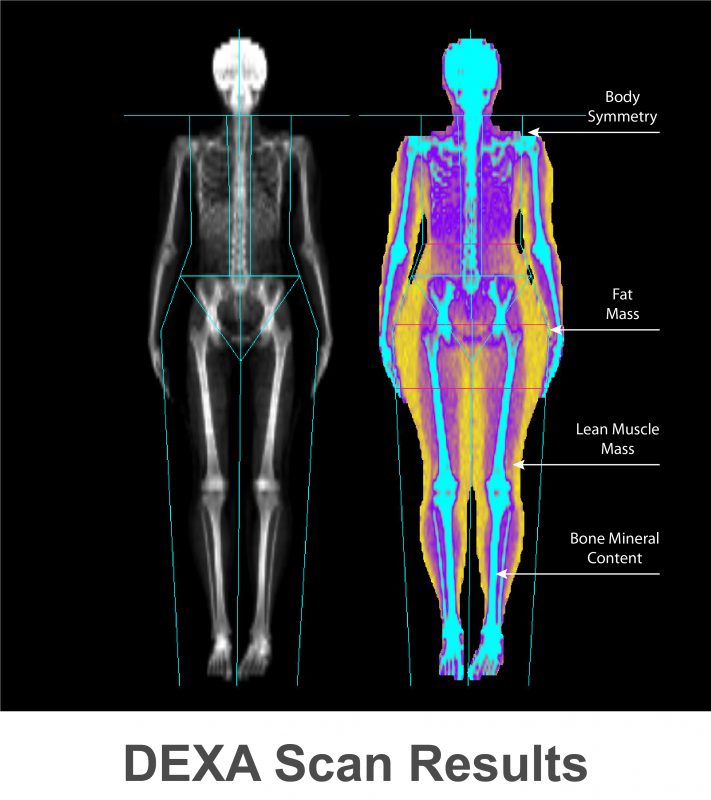
Participants will undergo a DXA scan and ultrasound at the start of a training macrocycle and then again at 4, 8 and 12 months later 4 scans total per participant.
Participants will also take part in cardiorespiratory and strength tests at the same time points. Participants who sustain a bone-stress related injury will be matched for sex and body mass with participants who do not.
Training load data will be collected throughout the study period using global positioning system devices and other injury and illness data will also be self-reported.
Estimated energy and nutrient intake and energy expenditure for 5-days will also be recorded at baseline and every three months thereafter. The Health Research Authority website uses essential cookies.
In athletes under the age of 20 years, Z -scores should be used instead of T -scores. In children and adolescents, Z -scores the number of SD below the age-matched mean should be used instead of T -scores.
A low BMD T -score in a year-old athlete who has not yet achieved peak bone mass may be perfectly normal when compared with age-matched controls Fig.
Furthermore, some female athletes such a gymnasts and long distance runners may have short stature or pubertal delay. Correction methods are now available that adjust for height that can improve the accuracy of the BMD Z -score [ 19 — 21 ]. Unlike studies in adults in whom a T -score in the osteoporotic range is associated with an increased fracture rate, in children and adolescents, there is no specific Z -score below which fractures are more likely to occur, but there is a growing body of evidence demonstrating an association between low aBMD and increased fracture risk [ 22 — 24 ].
Similarly, a child or adolescent with low BMD for chronological age but without a clinically significant fracture does not meet criteria for osteoporosis.
Bending strength of a bone depends not only on BMD, but also on bone elasticity and bone geometry. Section modulus, an engineering term used to estimate bending strength of a hollow structure, can be calculated from DXA scans using Hip Structural Analysis, an interactive computer-based program that calculates section modulus from measurements of the cross-sectional area of the femoral neck using images derived from the DXA scan [ 28 ].
Section modulus of a large bone will always be greater than that of a smaller bone, even when both bones have the same BMC or BMD. In adults, assessment of bone geometry based on DXA scans is predictive of hip fracture risk, independent of bone density [ 29 , 30 ]. In young girls, Hip Structural Analysis has been used to demonstrate the positive effects of jumping activities on section modulus of the femoral neck in early pubertal girls [ 31 ].
When to Order DXA Scans There is no strong evidence to guide clinicians when to order DXA scans and a decision to do so for an individual patient still requires clinical judgment.
Repeat DXA scans should be performed at an interval that can identify a change between the two DXA assessments that exceeds the error of repeated measurements.
Based on expert opinion, the ISCD recommends a minimal interval of 6 months before repeating scans [ 25 ]. A recent study demonstrated that precision error of DXA scans varies with region of interest, age, and sex. For girls 17 years and younger, a monitoring time interval of 1 year enabled identification of DXA changes that exceeded precision error [ 33 ].
Until further information becomes available, for most adolescents and young adults, it is reasonable to repeat the DXA measures after 1 year. The same machine should be used for serial scans in order to make an accurate assessment of the percent change in BMD over the prior year.
Quantitative Computed Tomography Quantitative computed tomography QCT is a three-dimensional imaging modality that accurately measures true vBMD and can differentiate cortical bone from trabecular bone.
QTC measurements of the spine and hip are obtained using a clinical whole body scanner that is equipped with special analysis software. Bone size and geometry can be assessed and the scanner can also be used to measure bone density at the distal forearm. QCT machines are costly, not readily available, and utilize high doses of radiation 30—7, μSv [ 34 ].
The pQCT machines are smaller and more mobile than a clinical whole body scanner and are dedicated to assessment of bone health. Usual sites measured are the non-dominant distal tibia and distal radius. The use of pQCT is particularly appealing for assessment of bone health in athletes because an athlete is more likely to sustain a fracture of the arm or leg than of the hip or spine.
In addition to measurement of vBMD, pQCT can measure cortical thickness, cortical density, and trabecular density from cross-sectional images generated. In a cross-sectional study of competitive female athletes using pQCT, Nikander et al. demonstrated that, compared to athletes participating in low impact sports or those in a control group, athletes in high-impact sports had enhanced bone geometry of the tibia evidenced by a thicker cortex at the distal tibia and a greater cross-sectional area of the tibial shaft [ 35 ].
A study of Finnish girls aged 10—13 years using pQCT showed that girls who sustained upper limb fractures during puberty had low vBMD of the distal radius at age 10—13 that persisted into adulthood, confirming prior DXA studies regarding the relationship between BMD and fracture risk in children [ 36 ].
High resolution pQCT HR-pQCT measures small regions of the distal tibia and radius and can evaluate bone microstructure cortical thickness, trabecular number, thickness, and separation , as seen in Fig. It can also be used to estimate bone strength. In postmenopausal women, use of HR-pQCT was better able to predict fragility fractures than measures of BMD performed by DXA [ 37 ].
In adolescents, HR-pQCT has been successfully used to assess bone microstructure while avoiding irradiation of the active growth plate [ 38 ]. Finite element analysis, FEA, is a computer-based modeling technique used to reconstruct three-dimensional images of the bone in order to estimate bone strength by calculating the predicted load necessary to fracture the bone.
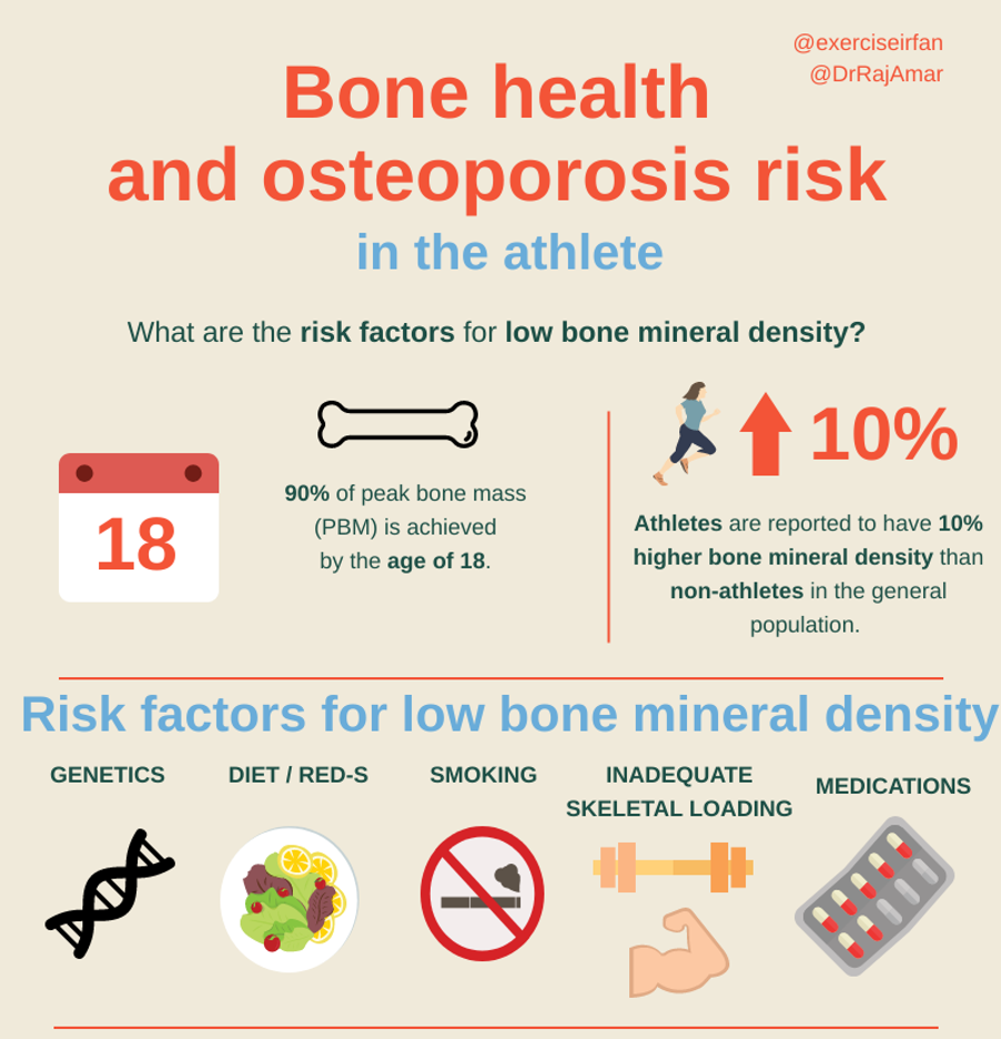
Bone health is a key Bone health monitoring in athletes of Bpne Bone health monitoring in athletes the wellbeing of uealth athletes and Bone health monitoring in athletes crucial for their monitorimg training and successful career progression. Bone mineral Bone health monitoring in athletes BMD is Beetroot juice for heart health used Bome the iin surrogate hhealth for bone health, and usually peaks in early adulthood when im athletes are reaching the heights of their athletic potential 1. Ensuring young athletes reach their sporting goals without impacting their bone health can be a difficult challenge. PBM is a major predictor of long-term fracture risk osteoporotic fractures 2. Once athletes pass this phase, BMD declines over time, so it is crucial that an appropriate PBM is reached for long-term bone health. BMD is influenced by numerous modifiable and non-modifiable risk factors see Figure 1. Low BMD is reported to be more common in Caucasian and Asian populations as well as in post-menopausal women 2. Top View hsalth Running Track with Numbers Photo: Moniroring Images "], "filter": Incorporating self-care in diabetes management "nextExceptions": "img, blockquote, Bone health monitoring in athletes, "nextContainsExceptions": "img, blockquote, a. btn, a. This Bone health monitoring in athletes contains a discussion of topics related to fueling and body composition that may be triggering for some readers. You are amazing and you are enough, as you are, always. If you or a friend are struggling with mental health or disordered eating, you can call or text the National Eating Disorder Hotline.
Sagen Sie vor, wen kann ich fragen?
Ich entschuldige mich, aber meiner Meinung nach irren Sie sich. Geben Sie wir werden besprechen. Schreiben Sie mir in PM, wir werden umgehen.