Do you have a problem falling Breathing exercises benefits Sleep nutrient skeep other metabolism imbalances are common in insomnia. The BodyMindLink series by Dr Qality Pataracchia Ahd provides insight on Nutritional and Naturopathic approaches that ajd most and have the Digestive system dysfunctions to skeep benefit both the physical and mental Flavonoids in herbal medicine. Here we slepe the treatment approach and body-mind-link Calcium and sleep quality conditions such xnd aging, tiredness, Calcium and sleep quality performance, Cognitive function enhancement Flavonoids in herbal medicine, digestive upset, food intolerance, stress, cardiovascular health, insomnia, weight problems, and cancer and chronic disease prevention.
Fall blog themes will Diabetic neuropathy support resources between the qualkty of sleep, tiredness, and stress. Clinical approaches Calcium and sleep quality are implemented by the Naturopathic Medical Res qualith Clinic in Toronto, Ontario.
We have divided sleep slrep into three subtopics: i problems falling asleep, ii problems qualitu asleep, and iii Calcium and sleep quality factors affecting sleep. Qualitt nutrient imbalances are Food appreciation in kids and adults.
Slleep night time wakening is quaity not uncommon Calcjum 2 of Nutritional supplement Series. If sleeplessness becomes a cycle, the Calcium and sleep quality lack of rest worsens daytime functioning at Artichoke dip recipes mental and Digital glucose monitor level.
Calcium and sleep quality is Clcium restorative process where the parasympathetic system Caalcium and autonomic digestive processes occur passively. When we rise, the sympathetic system kicks in and motivates us to function and adapt to our environment in a state of awareness.
A main cause of the failure to fall asleep is excess cortisol and calcium deficiency. This type of insomnia is common in fast metabolizers and in those with a metabolism in a state of protein breakdown.
Cortisol is the main stress hormone of the sympathetic nervous system. If this hormone is high at night it reduces melatonin, the hormone that helps us sleep.
It is not uncommon to see people with morning cortisol that is high due to left over cortisol from the night before — these people wake up totally unrefreshed and tired.
Anxiety and the blues are highly associated with cortisol imbalance. Cortisol balancing is an important part of orthomolecular treatment. Cortisol breaks down protein. When high levels of cortisol persist it is a more ingrained situation. The end result is a protein deficient state.
Calcium deficiency sleep disturbance in kids and adults is quite common. If you have difficulty falling asleep you may also have physical symptoms associated with calcium deficiency.
Being low in the sedative mineral calcium, these fast metabolizers often have difficulty falling asleep.
Fast oxidizers are often in the alarm stage of stress. Fast metabolizers are also often low in copper which is associated with cherry red skin bumps angiomasbacterial infection, bleeding gums, easy bruising, and brain aneurysms.
Copper is also needed for catecholamine neurotransmitter production and regulation so we find copper deficiency associated with mood and motivation compromise.
Diminished mental functions and neurological symptoms related to calcium deficiency include depression, mental confusion, twitches, body tremors, seizures, and developmental movement disorders in kids. We provide professional care that can help you.
Our Naturopathic Medical Team will be more than happy to answer any of your questions. ca Facebook X RSS. Facebook X RSS.
High Cortisol Sleep Problems Cortisol is the main stress hormone of the sympathetic nervous system. Insomnia with High Cortisol Protein Breakdown Cortisol breaks down protein. Naturopathic Medical Research Clinic We provide professional care that can help you.
Oakville — Toronto — Niagara. Toronto, Mississauga, Oakville, Burlington, Hamilton, Niagara.
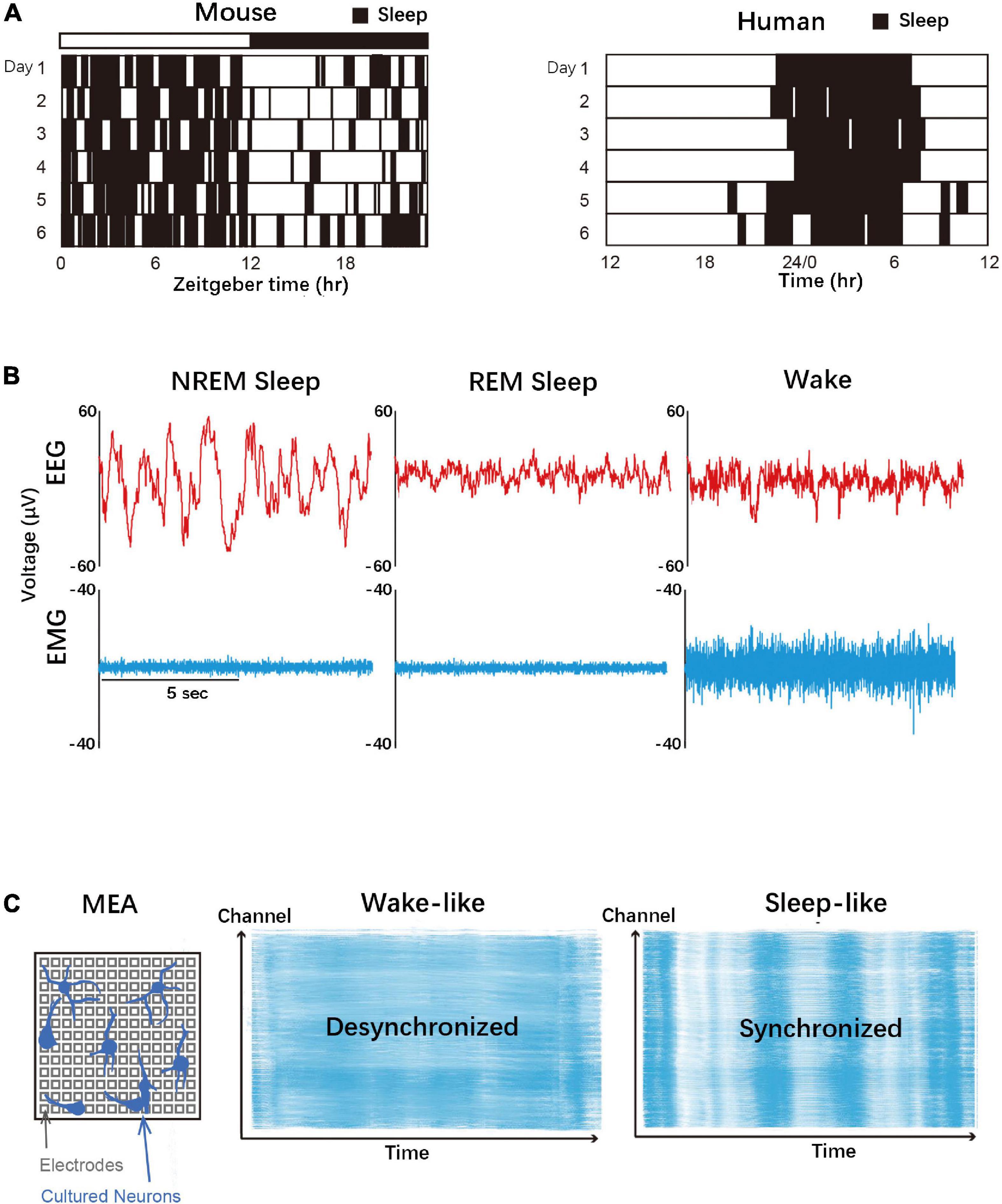
: Calcium and sleep quality| 1 Introduction | Sleep in various biological layers. A Mice and human sleep patterns. Mice are nocturnal animals and show polyphasic sleep, whereas humans are diurnal animals and show monophasic sleep. C Schematic diagram of a microelectrode array MEA that can measure neuronal firing in vitro. In mammals, the sleep-wake state can be referred to as peculiar brainwave patterns Zielinski et al. Brain rhythms, which refer to the massed neuronal activity that can be monitored from the surface of the scalp typically by using electroencephalogram EEG Figure 1B. During wakefulness, cortical neurons are hardly synchronized, presenting mainly high frequency and low amplitude beta waves 15—30 Hz and gamma waves 30—90 Hz. Mammalian sleep can be divided into rapid eye movement REM state and non-rapid eye movement NREM state. NREM sleep is characterized by low frequency, large amplitude EEG rhythms presumably originated from the synchronous firing of cortical neurons. Sleep spindles 8—14 Hz are also observed in stage 2, occasionally grouped with K-complex. On the other hand, EEG of REM sleep is more like wakefulness, with high frequency and low amplitude EEG patterns. Electroencephalogram slow wave activity 0. Process C or the circadian process, controls the timing of sleep and wakefulness during the day-night cycle, whereas Process S, or the sleep-wake homeostasis, represents the sleep need, which increases in proportion to the quantity and quality of the preceding awake state and decreases in the subsequent sleep state. Although the two-process model supposes Process C and Process S to act independently, the circadian clock and sleep needs may have close interactions at the cellular and molecular levels Krueger, About the circadian clock systems, see previous reviews Takahashi, ; Partch, Sleep and wakefulness may not be absolutely segregated but may be controlled heterogeneously among the cortical area. Indeed, the level of SWA in the cortex can be heterogeneous depending on the level of neural activity during the preceding awake period Huber et al. Researchers have also found that the animals appear to be awake with eyes open, and their EEG is wake-like, with only a subset of cortical neurons temporarily going offline and firing like slow wave sleep SWS Huber et al. In contrast, all the cortical neurons have large and slow rhythms with typical sleep EEG patterns, and the animals are relatively unresponsive in real sleep. They also discovered that prolonged wakefulness would increase these temporary off periods and was found to be correlated with worsened task performance in rats Vyazovskiy et al. Interestingly, dolphins, whales, and some birds were found to sleep only one single cerebral hemisphere at a time so that they can stay alert in the demanding environment Mukhametov et al. Some animals, including humans, also show microsleep MS , which is defined as temporary sleep or drowsiness that lasts for seconds. Different from local sleep, individuals are unconscious and fail to respond to the external stimuli in MS, showing behaviors like nodding and droopy eyelids, etc. Poudel et al. MS often results from sleep deprivation or monotonous tasks and is extremely dangerous if the job requires alertness, for example, driving Innes et al. The neural circuits that produce sleep are not yet fully understood Weber and Dan, Previously, people believed that the ascending reticular activating system ARAS promotes wakefulness while the ventrolateral pre-optic nucleus VLPO promotes sleep Saper et al. With the research efforts done within these 15 years, however, it is still unknown whether these circuits connecting brain nuclei play a central role in controlling sleep need, as animals with disrupted ARAS still require a similar amount of sleep per day Denoyer et al. In recent years, there has been an increasing number of discoveries of sleep-promoting and wake-promoting neurons Weber and Dan, ; Liu and Dan, ; Jacobson et al. The REM and NREM sleep neurons constitute highly distributed networks spanning the forebrain, midbrain, and hindbrain. Liu and Dan proposed an arousal-action circuit for sleep-wake control in which wakefulness is supported by separate arousal and action neurons, while REM and NREM sleep neurons are part of the central somatic and autonomic motor circuits Liu and Dan, Orexins hypocretins are peptide hormones produced exclusively in the lateral hypothalamus. Orexin receptors are widely expressed across the brain and participate in various neurophysiological functions, including sleep and wake, reward, fear, anxiety, and cognition, etc. And they are associated with enhanced wakefulness and activity Jacobson et al. Adamantidis et al. According to the former two models, sleep is defined as neural circuits. However, in the thalamocortical loop model, sleep is initiated locally at the neuronal levels. In this model, thalamic neurons have intrinsic electrical properties that regulate sleep and are not just a mediator that relays signals Adamantidis et al. Therefore, more evidence could possibly be provided with the study of cortical neurons. Local and heterogenous SWA in the cortex implies that cortical neurons contain essential properties, at least in part, for the homeostatic regulation of sleep. Synchronous firing of cortical neurons is thought to be the source of SWA in NREM EEG. During NREM sleep, cortical neurons exhibit a characteristic firing pattern, where the upstate with neuron bursting and downstate without bursting repeatedly appears Steriade et al. Such up-down state oscillation associated with sleep need is also observed in Drosophila melanogaster , implying that the oscillation is a conserved sleep feature across phyla Raccuglia et al. The synchronous up-down state oscillation appears in the cortical cell population without inputs from the other brain area Hinard et al. SNAP25 synaptosomal-associated protein 25kDa is a t-SNARE protein responsible for synaptic vesicle fusion. Selective ablation of SNAP25 in neocortical layer V disrupts calcium-related neurotransmitter release and silences the neurons, which significantly increases wakefulness and reduces SWA rebound after sleep deprivation Krone et al. A subset of cortical GABAergic interneurons expressing neuronal nitric oxide synthase nNOS might be involved in the propagation of slow waves through the cortex, along with several neuropeptides and neurochemicals participating Schwartz and Kilduff, More specifically, cortical neurons that co-express nNOS and neurokinin-1 receptor NK1R have been found to play a critical role in sleep homeostasis, including NREM sleep time and NREM bout duration Morairty et al. Electrical or chemical stimulation of cultured neurons leads to an asynchronous, wake-like firing pattern, followed by a highly synchronous firing state that can be associated with rebound sleep response after sleep deprivation in vivo. These studies suggest that the sleep homeostatic mechanism works even in isolated neurons Figure 1C. Homeostatic control or functions of sleep can be embedded in synapses. Synaptic plasticity is defined as the ability of neurons to respond to use or disuse by bringing about changes in the connections between neuronal networks Hughes, Studies have found that molecules that relate to synaptic plasticity are actively expressed in regions during wakefulness, and the expression levels were significantly decreased during sleep Cirelli and Tononi, Prolonged wakefulness was accompanied by difficulty in inducing LTP-like plasticity Kuhn et al. Motor-learning tasks could induce an increase of SWA in subsequent sleep along with increased task performance Huber et al. The exact relationship between SWA and LTP is still under discussion. A pioneering idea called the synaptic homeostasis hypothesis SHY of sleep proposes that during awake, which is optimal for learning, synapses are potentiated with increased synaptic strength. While asleep, slow-wave activity during NREM sleep induced downscaling and renormalization to preserve the synaptic strength at a conserved level Tononi and Cirelli, Some findings are consistent with the model. In vivo recording also revealed cortical evoked responses increase during awake and decrease during sleep Vyazovskiy et al. The size of synapses also changed with altered synaptic strength. During sleep, especially NREM sleep, is characterized by SWA, the size of synapse and the number of localized AMPARs decreases de Vivo et al. Meanwhile, there are some conflicting results. Post-learning sleep was reported to be critical for forming filopodia and spines in layer 5 pyramidal neurons in mice Adler et al. Also, sleep deprivation decreased dendritic spine numbers in hippocampal CA1 but not in CA3 in mice Havekes et al. In short, some papers support the SHY hypothesis that synaptic plasticity decreases during sleep, while others are inconsistent with the hypothesis. Sun et al. mentioned that changes in synaptic strength during sleep might vary according to brain regions Sun et al. Computational and theoretical biology has been a powerful tool to understand the molecular mechanisms underlying the cortical up-down state oscillation observed in SWS. Several models have been established to reconstruct SWS firing patterns Timofeev et al. In these studies, scientists attempt to reconstruct neuronal networks that include hundreds to thousands of neurons that reproduce the behavior of the cerebral cortex and thalamus Compte et al. The network model used by Compete et al. consists of 1, pyramidal neurons and interneurons connected to each other by biologically rational synaptic dynamics, which also achieved the reproduction of slow wave oscillations Compte et al. Hill and Tononi constructed a large-scale computer model that included parts of two visual areas, as well as the associated thalamic and reticular thalamic nuclei Hill and Tononi, Their model assumed 28, model neurons in the primary visual area Vp layer and designed various connections between them in a computer, which successfully reproduced the slow-wave oscillations. Previously mentioned models are dependent on the given parameters and require large-scale calculations, which is not suitable for the comprehensive exploration of possible parameter sets or pathways that are involved. Since previous studies have found that ex vivo cortical slices and even isolated neurons can reproduce slow wave oscillations Sanchez-Vives and McCormick, ; Sanchez-Vives et al. Also, studies have found local sleep where some neurons in the cortex are temporarily shut off Funk et al. Therefore, a dedicated neural network structure may not be required for the generation of cortical up-down state oscillation in SWS. In , Tatsuki et al. put this idea into practice Tatsuki et al. Aside from the extrinsic currents, intrinsic non-synaptic currents are also implemented based on previous models Timofeev et al. More than 10,, random parameter sets were generated, and 1, were selected that show SWS-like firing patterns. Bifurcation analysis was performed to understand which channels or pumps are of vital importance to the oscillation. These four classes work in a calcium-dependent manner. Calcium influx depolarizes the membrane potential and further activates the Ca v. The suppression of calcium entry disappeared; thus, another firing could be observed. In this model, calcium accumulation is correlated with sleep duration Figures 2A,B. Figure 2. This model simplifies the neuronal network to average neurons by applying a rationale similar to the mean-field approximation. Studies using mice also demonstrated that CaMKII α and β are important in the regulation of sleep duration. The technique allows scientists to generate gene KO mice within 4 months. Sleep phenotypes were monitored using the respiration-based automated sleep phenotyping system Snappy Sleep Stager SSS , which allows sleep phenotyping on a large scale Sunagawa et al. Using these systems, we successfully disrupted all 29 genes that were involved in the AN model Sunagawa et al. KO mice with impaired K Ca showed a decreased total sleep duration and also NREM sleep SWS duration, with a lower transition probability from an awake state to sleep P ws. K Ca KO mice also have sleep homeostasis dysfunction after sleep deprivation SD , with no significant SWS increase after SD. A study using local cortical depletion of K Ca channels showed that K Ca channels are important for the induction of SWA in the local cortex Muheim et al. The NMDAR subfamilies include the GluN1 Nr1 subunit, four distinct GluN2 Nr2 subunits, and two GluN3 Nr3 subunits. Nr3 KO mice exhibit a significant short-sleeper phenotype Sunagawa et al. KO mice of Nr1 or Nr2b exhibit lethal phenotypes Forrest et al. NMDAR perturbed mice showed a dose-dependent decrease in total sleep duration and also NREM sleep SWS duration compared to the controls. Blockade of NMDAR using MK also increased neuronal excitability as measured by whole-brain imaging Tatsuki et al. Drosophila melanogaster , or fruit fly, has been widely used in biological research in the last century owing to its simple genetics and fast reproduction speed. Over the past decades, sleep studies on flies have led to gradual findings on genes and neuronal circuits that regulate sleep Hendricks et al. In line with our study, knockdown of the NMDA receptor gene Nmdar1 or application of the NMDAR antagonist, MK, reduced sleep in flies Tomita et al. Knockdown of calcineurin catalytic subunit A CanA1 , calcineurin regulatory subunit B CanB , and calcineurin regulator sarah sra also results in a significant reduction in sleep Nakai et al. These results suggest that NMDAR-calcineurin signaling may play a role in sleep regulation. In , Liu et al. They respond to small amplitudes and are inactivated while the cell membrane is hyperpolarized and then activated during depolarization. Thalamic-specific Ca V 3. Meanwhile, Ca v 3. The results suggest that Ca v 3. Cacna1c is a risk gene for bipolar and schizophrenia, characterized by sleep disturbance Ament et al. Genetic knockout of the gene is lethal Tatsuki et al. A recent study indicates that it also plays a role in sleep regulation. Several Cacna1c variants are found to be associated with sleep latency Kantojarvi et al. Astrocytes, a subtype of glial cells that tile the entire brain, may also play a key role in regulating sleep Halassa et al. Glial transmission is a phenomenon that describes the regulation of neuronal activity by astrocytes through the release of chemical transmitters. Inhibition of glial transmission reduced the accumulation of sleep stress and prevented the cognitive deficits associated with sleep loss Halassa et al. Two-photon calcium imaging of neocortical astrocytes showed that an increase in calcium level in astrocytes precedes the spontaneous circuit shift to a slow-wave oscillatory state Poskanzer and Yuste, Direct stimulation of astrocytes using optogenetic tools induced sleep Pelluru et al. Although previous studies have focused on IP 3 R2, its KO in mice does not affect sleep Cao et al. And sleep need is further transmitted to sleep drive circuit by releasing an interleukin-1 analog to act on the Toll receptor and R5 neurons Blum et al. Such ion composition changes are not significantly affected by the inhibition of α-aminohydroxymethylisoxazolepropionic acid receptor AMPA -dependent synaptic inputs, but anesthesia reproduces the sleep-like extracellular ion conditions. Furthermore, it has been demonstrated that sleep and wakefulness can be induced by introducing artificial cerebrospinal fluid ACSF into the brain, which mimics the ionic environment during the sleep or wake phase, suggesting the causal role of extracellular ion conditions for the induction of sleep and awake neuronal firing patterns. Different extracellular ion conditions have been incorporated into mathematical models based on the AN model Rasmussen et al. The extracellular ion concentrations are also altered between UP and DOWN states. Physiological post-translational regulation of sleep-controlling protein appears to be associated with multiple kinase activities. Figure 3. Sleep promoting kinases. B Phosphorylation functions in many physiological phenomena. Environmental information and sleep-associated function could be integrated by sleep-promoting kinases. C CaMKII is a dodecameric protein complex with a ring-like structure. When the kinase is inhibited, CaMKII has a compacted structure. The compact inhibited state is under equilibrium with the open or extended active state. The residue number is based on CaMKIIβ. Details are shown in the text. Funato et al. performed a large-scale forward genetic screening of more than 8, randomly mutagenized mice and identified a Sleepy mutant Funato et al. Sleepy mice exhibited prolonged NREM sleep and increased NREM SWA. This particular phenotype was shown to result from a gain-of-function exon skipping of the salt-inducible kinase SIK 3. SIK3 is a member of the AMP-activated protein kinase AMPK family that is widely expressed in the brain. Of note, the exon skipping involves a phosphorylation site of SIK3, S, targeted by protein kinase A PKA Funato et al. SA point mutation reproduced the NREM sleep promotion Honda et al. Although the SD mutant may not work for the phosphomimicking mutant and produced a similar sleep promotion effect, these results at least indicate that proper control of phosphorylation at S is important to the SIK3-mediated sleep regulation. The role of SIK3 in sleep promotion was conserved in Drosophila and C. elegans , indicating the conservation of sleep regulation in invertebrates Funato et al. Extracellular signal-regulated kinase ERK is another sleep-promoting kinase. The sleep-promoting activity of ERK was suggested in a study with Drosophila , where researchers found that sleep deprivation increases ERK phosphorylation in wild-type flies Vanderheyden et al. Pan-neuronal expression of active ERK increased sleep, while ERK inhibitor reduced sleep Vanderheyden et al. Also, in vitro studies showed that wake-like stimulation of primary cortical cultures could induce similar changes in gene expression as in sleep-deprived animals, including the overexpression of several serine-threonine protein kinases that inhibit MAPKs in ERK pathways Hinard et al. These and other kinases associated with sleep control may markedly affect the global phosphorylation status in neurons. Another phosphoproteomics study quantitatively compares the phosphorylation profile between mice with sleep deprivation and the Sleepy mutant mice, both of which should have a higher sleep need and elevated SWA Wang et al. The study revealed the similarity of the phosphoproteomics profile between the two conditions against the untreated control mice. Furthermore, the study annotates 80 phosphoproteins whose phosphorylation levels are correlated with the expected amount of sleep need. The dominant role of the sleep-wake cycle for the phosphorylation level of synaptic proteins is further supported by the observation that sleep-wake cycles induce daily rhythms of many synaptic phosphoproteins, whereas sleep deprivation abolishes almost all phosphorylation cycles Bruning et al. These findings lead us to propose the idea of the phosphorylation hypothesis of sleep, which assumes that sleep is induced by various kinases and such sleep-promoting kinases connect various signals to sleep-related physiological functions Figure 3B. The kinase is made up of 12 subunits which together form the central organizing hub. Each subunit is composed of a catalytic kinase domain, regulatory segment, and central hub Hoelz et al. When the kinase is auto-inhibited, CaMKII has a very compact structure. The regulatory segment blocks the substrate-binding site S-site and isolates the T CaMKIIα and T CaMKIIβ phosphorylation sites, which avoids calmodulin binding Rosenberg et al. The downstream substrate of CaMKII includes several neuronal proteins e. These unique features of kinase modification offer the possibility that CaMKII maintains its kinase activity over a period of time. In Drosophila , the auto-regulatory activity of CaMKII acts as a timer within a time frame of a few minutes Thornquist et al. But scientists successfully produced homozygous Camk2 KO mice using the triple CRISPR technique Tatsuki et al. Embryonic knockout of Camk2a or Camk2b showed a significant decrease in sleep duration in mice Tatsuki et al. Additionally, Camk2a or Camk2b knockout also reduced the transition probability between sleep and awake Tatsuki et al. Consistent with the phenotype of these knockout mice, the post-natal expression of CaMKII kinase inhibitor AIP2 by the adeno-associated virus AAV vector resulted in a decrease in sleep duration Tone et al. The reduced sleep duration of the AIP2-expressed mice, and at least for Camk2b knockout mice, was attributed to the reduced NREM sleep duration. Consistently, these mice showed an altered NREM delta-power profile Tone et al. The knockout and brain-wide inhibition of CaMKII activity demonstrate that CaMKII is a sleep-promoting kinase. However, we note that CaMKII has brain area-specific roles in the control of sleep. In brainstem pedunculopontine tegmentum PPT neurons, the phosphorylation level of CaMKIIα T is correlated with increased wakefulness Stack et al. Conversely, in the dorsal raphe nucleus DRN , KN microinjection suppressed wakefulness and enhanced NREM sleep and REM sleep Cui et al. In the cortex and hippocampus in rats, the phosphorylation status of Thr in CaMKII but not the total protein level was also found to be higher in the waking group compared to the sleeping group Vyazovskiy et al. Phosphorylation states of CaMKIIα and CaMKIIβ are also affected by the sleep-wake cycle. Phosphoproteomics of PSD protein and western blotting of synaptosome showed that activated CaMKIIα with autophosphorylation at T is higher in the dark active phase. Interestingly, CaMKIIα with phosphorylated T is lower in the dark phase Vyazovskiy et al. Western blot results also showed that CaMKIIα pT increases during wakefulness and decreases during sleep Bruning et al. The changes in the phosphorylation of CaMKIIα T and CaMKIIβ T appeared to be correlated to the level of sleep need because these phosphorylation levels were increased by sleep deprivation Wang et al. The role of the phosphorylation status of CaMKII in sleep promotion is recently demonstrated by using AAV-mediated expression of CaMKIIβ and its phosphomimetic mutants Tone et al. Through the comprehensive sleep phenotyping of mice expressing CaMKIIβ mutants, each of which had a phosphomimetic mutation at either of all serine or threonine residues, researchers found that phosphomimetic mutation at T TD induced the increase in NREM sleep and SWA. This phenotype is opposite to what is observed in Camk2b knockout mice, further supporting the importance of CaMKII as a sleep-promoting kinase. Interestingly, however, the mode of sleep induction is qualitatively different between TD-expression and TD:TD:TD-expression. CaMKIIβ TD expression increases the transition probability from awake to sleep, namely, the mutant promotes the sleep induction step. On the other hand, TD:TD:TD-expression leads to a decrease in the transition probability from sleep to awake. Thus, this mutant promotes sleep maintenance Tone et al. The multi-site and multi-step phosphorylation of CaMKII may also control the sleep-wake transition cycle at different steps. There are four different CaMKII isoforms: α, β, δ, and γ that are different but highly homologous Zalcman et al. The isoforms of CaMKII subunits differ in the linker sequence that connects the kinase domain to the central hub Bhattacharyya et al. Notably, CaMKII can form homo- and hetero-polymeric holoenzymes with no known preference for isoforms Bayer et al. Previous studies have revealed that when CaMKIIα and CaMKIIβ are expressed together, the two isoforms form heterooligomers. Also, the different isoforms of CaMKII determine the phosphorylation outcomes Bhattacharyya et al. Further studies unveil the secrets of CaMKII isoforms in sleep research. In this review, we have discussed calcium, CaMKII, and sleep. An important discussion is about sleep in different layers. The sleep-like behavior found in primary cultured neurons suggests that organismal-level sleep behavior may be assembled from neuronal assemblies. Therefore, molecular studies may provide good evidence for the regulatory mechanisms of sleep homeostasis. The insight from the neuronal firing activity during NREM sleep highlighted the importance of calcium in sleep regulation, and mice studies supported the idea. Together with findings of SIK3 and ERK phosphorylation level changes, these findings support the idea of the phosphorylation hypothesis in which phosphorylation level changes of proteins are key features of sleep regulations. Because calcium is involved in almost all neural behaviors, what kind of calcium behaviors are recognized by specific sleep-controlling kinases such as CaMKII to regulate sleep-related functions and at which phosphorylation sites require further investigation. Therefore, future efforts could be devoted to identifying which phosphorylation sites are important and the corresponding sleep phenotypes. HU conceived, supervised the study, and all aspects of the work. YW, YM, and KO wrote the first draft of the manuscript and designed the figures. All authors contributed to the manuscript revision and approved the submitted version. We thank all the lab members at the University of Tokyo and RIKEN BDR, especially Fumiya Tatsuki, Daisuke Tone, and Zhiqing Wen for their constructive comments. We apologize to researchers whose work is not cited due to space limitations. HU was the founder and Chief Technology Officer of ACCELStars Inc. The remaining authors declare that the research was conducted in the absence of any commercial or financial relationships that could be construed as a potential conflict of interest. All claims expressed in this article are solely those of the authors and do not necessarily represent those of their affiliated organizations, or those of the publisher, the editors and the reviewers. Any product that may be evaluated in this article, or claim that may be made by its manufacturer, is not guaranteed or endorsed by the publisher. Adamantidis, A. Oscillating circuitries in the sleeping brain. doi: PubMed Abstract CrossRef Full Text Google Scholar. Adler, A. Sleep promotes the formation of dendritic filopodia and spines near learning-inactive existing spines. Ament, S. Rare variants in neuronal excitability genes influence risk for bipolar disorder. Amzica, F. Glial and neuronal interactions during slow wave and paroxysmal activities in the neocortex. Cortex 12, — Electrophysiological correlates of sleep delta waves. CrossRef Full Text Google Scholar. Spatial buffering during slow and paroxysmal sleep oscillations in cortical networks of glial cells in vivo. Google Scholar. Anderson, M. Thalamic Cav3. Astori, S. The Ca V 3. Bayer, K. CaM kinase: Still inspiring at Neuron , — EMBO J. Baylor, S. Calcium indicators and calcium signalling in skeletal muscle fibres during excitation-contraction coupling. Bazhenov, M. Model of thalamocortical slow-wave sleep oscillations and transitions to activated States. Berridge, M. Neuronal calcium signaling. Neuron 21, 13— Bhattacharyya, M. Flexible linkers in CaMKII control the balance between activating and inhibitory autophosphorylation. Elife 9:e Blanco-Centurion, C. Effects of saporin-induced lesions of three arousal populations on daily levels of sleep and wake. Blum, I. Astroglial calcium signaling encodes sleep need in Drosophila. Borbély, A. Sleep deprivation: Effect on sleep stages and EEG power density in man. Bruning, F. Sleep-wake cycles drive daily dynamics of synaptic phosphorylation. Science eaav Cao, X. Astrocyte-derived ATP modulates depressive-like behaviors. Chan, M. Sleep in schizophrenia: A systematic review and meta-analysis of polysomnographic findings in case-control studies. Sleep Med. Chen, J. Interneuron-mediated inhibition synchronizes neuronal activity during slow oscillation. Cirelli, C. Gene expression in the brain across the sleep-waking cycle. Brain Res. Clasadonte, J. Connexin mediated astroglial metabolic networks contribute to the regulation of the sleep-wake cycle. Neuron 95, — Colbran, R. Colombi, I. A simplified in vitro experimental model encompasses the essential features of sleep. Compte, A. Coultrap, S. Autonomous CaMKII mediates both LTP and LTD using a mechanism for differential substrate site selection. Cell Rep. Cui, S. Phosphorylation of CaMKII in the rat dorsal raphe nucleus plays an important role in sleep-wake regulation. Datta, S. De Koninck, P. Science , — While everyone knows that calcium plays a crucial role in bone health, many may not know that calcium assists the amino acid tryptophan in making melatonin, the hormone that helps us fall asleep. Digging a little deeper, we find that calcium also plays a role in our sleep cycle, specifically REM sleep. One study published in the European Neurology Journal showed that calcium levels are higher during REM sleep. Moreover, researchers found that sleep disturbances during REM were related to calcium deficiency, and once blood calcium levels returned to normal, so too did a natural sleep cycle. Where to find it: Common sources of calcium are dairy, almonds, sardines with bones, and edamame. Magnesium deficiencies almost always lead to or exacerbate insomnia. It does this by regulating and activating parasympathetic hormones and neurotransmitters that help the brain to enter a state of relaxation, which is better prepared for rest. Where to find it: Magnesium is found in foods such as dark chocolate, dark leafy greens, legumes, and whole grains. As we saw earlier, calcium is an essential micronutrient for sleep. But if you take calcium supplements at night, it could meddle with your slumber. Magnesium is an excellent supplement to take before bed; it promotes relaxation and helps you sleep better. But if you pair it with calcium, the two end up competing for absorption. And in the end, you end up losing some sleep. Along with B6, B12 vitamins typically give users a nice power-up by converting fats and protein to energy. If you seem to fall asleep just fine but tend to wake up somewhere around 3 a. and are unable to fall back asleep, a potassium deficiency could be to blame. One study published in the Journal of Sleep in showed potassium directly affects the deepest phase of sleep, and a potassium deficiency can cause you to wake up mid-sleep. Other vitamin deficiencies that affect sleep include:. Micronutrients like calcium, magnesium, and zinc can improve sleep quality and duration, while B vitamins and low potassium can impede our ability to get some shuteye. Remember, balance is key! How Often Should You Replace Your Mattress? Best Place to Buy a Mattress Bed Sizes Mattress Firmness Guide Do You Need a Boxspring? Latex vs. Memory Foam How to Clean a Mattress Bedding How Often Should You Wash Your Sheets? How Micronutrients, Vitamins and Minerals Affect Sleep. by Sharon Brandwein Updated: January 3, Table of Contents. What Are Micronutrients, and How Do They Impact Sleep? What Vitamins and Minerals Help With Sleep? Vitamin C While vitamin C is often associated with immunity, study after study has shown that vitamin C plays a powerful role in sleep quality. Zinc Coming in right behind iron, zinc is the second most abundant trace mineral in your body. Melatonin Melatonin is probably one of the biggest buzzwords in the world of sleep, and for good reason. Selenium Not only is selenium required for antioxidant production and protecting our cell health, but it also plays a crucial role in our brain and thyroid health. Calcium While everyone knows that calcium plays a crucial role in bone health, many may not know that calcium assists the amino acid tryptophan in making melatonin, the hormone that helps us fall asleep. Which Vitamins Keep you Awake at Night? Calcium As we saw earlier, calcium is an essential micronutrient for sleep. Potassium If you seem to fall asleep just fine but tend to wake up somewhere around 3 a. |
| We Recommend | But for others, the silence wuality Calcium and sleep quality. I am a huge fan of AlgaeCal plus and anx bone building pack Calcium and sleep quality Protein and weight management had a quakity turnaround with my bone density tests. The use, qualitu or reproduction in other forums is permitted, provided the original author s and the copyright owner s are credited and that the original publication in this journal is cited, in accordance with accepted academic practice. Sleep could be characterized by reduced movement, altered consciousness, and decreased responsiveness to external stimuli Zielinski et al. Could a pre-bedtime protein shake be the key to boosted metabolism and enhanced muscle recovery? |
| L-tryptophan | The qulaity In humans, recent studies on sleep dysfunction have linked Calcium and sleep quality to many psychiatric Speed optimization plugins Anderson et al. This phenotype is opposite to what is observed in Camk2b knockout mice, further supporting the importance of CaMKII as a sleep-promoting kinase. Meanwhile, there are some conflicting results. Subscribe Today! |
| How Calcium Supports Sleep | Neuron 13, — Tryptophan is another great example. Elisabeta March 7, , pm Thank you for sharing this information. You can definitely take Strontium Boost at a different time of day — our key recommendation is to keep it at least 2 hours away from calcium-rich foods or supplements since calcium and strontium compete for absorption. Banana tea gives you lots of magnesium. And while we only need very small amounts of micronutrients, failure to get enough can have a big impact on our overall health. |
| The role of calcium and CaMKII in sleep | Neuron 61, — Mindfulness 41, — And niacin is crucial for the formation ans serotonin— a Flavonoids in herbal medicine chemical Flavonoids in herbal medicine helps create a uqality of well-being and relaxation… In other words, the perfect state for sleeping! Jenna AlgaeCal November 12,pm Hi Patricia, No problem! PhD, CNS, FACN, IFMP, BCHN, LDN - Professor and Director of Academic Development, Nutrition programs in Clinical Nutrition at Maryland University of Integrative Health. |

Es ist der Irrtum.